Pelvic Inflammatory Disease Ultrasound Images
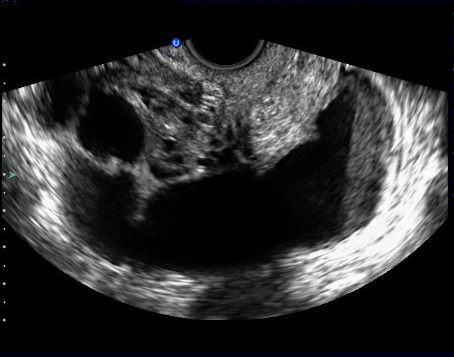
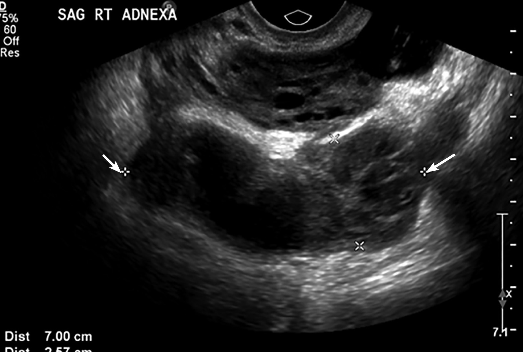
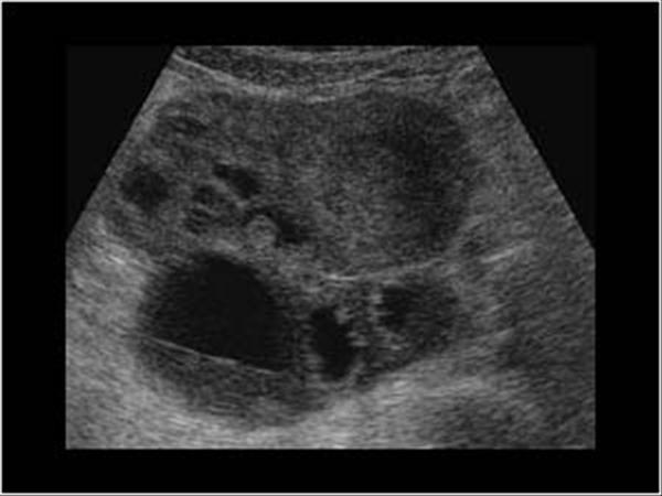
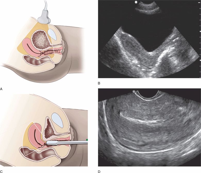
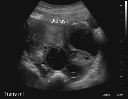
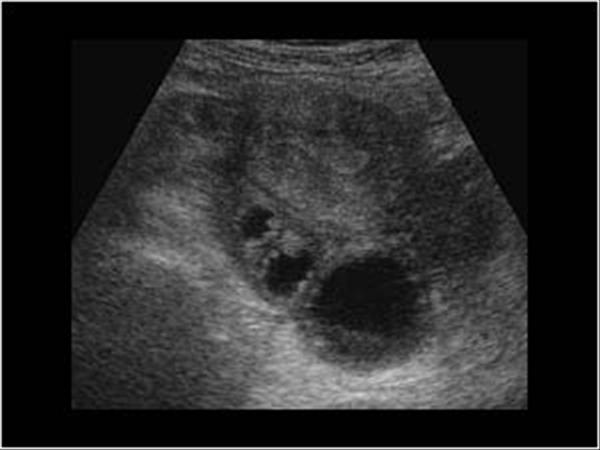
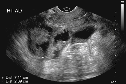
Pelvic inflammatory disease ultrasound images. 20 21-year-old woman with pelvic inflammatory disease. Though the study may be normal or sometimes non-specific there are a variety of findings that are characteristic of this process. Pelvic sonography is commonly performed in patients with a clinical diagnosis of pelvic inflammatory disease.
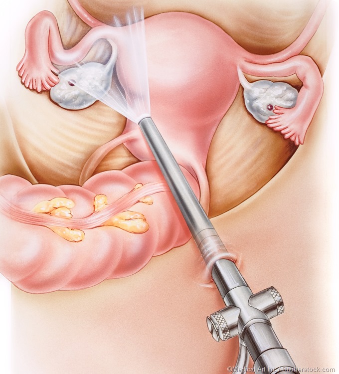
The doctor probes the lower abdomen in a girl who has pain and inflammation of the bladder. Pelvic ultrasound to provide images of the internal organs in the pelvic area. Biopsy which involves taking small tissue samples from the uterus.
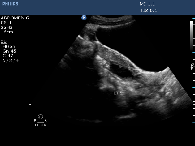
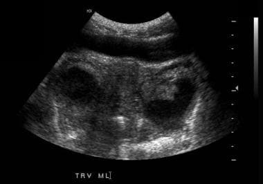
The sonographic markers of tubal inflammatory andor pelvic disease are summarized in Figure 7. Causes of Pelvic Inflammatory Disease. Transabdominal image of the right pelvis in a patient with pelvic inflammatory disease demonstrates dilated loops of small bowel without peristalsis compatible with a reactive ileus.
Although the study may be normal or sometimes nonspecific there are various findings that are characteristic of this process. The sonographic images are classified as acute and chronic and in each of these categories there is a section depicting wall thickness incomplete septa and wall structure. Female reproductive system diseases and treatment concept bladder inflammation pelvic inflammatory disease stock pictures royalty-free photos images.
Understanding of the sonographic features of pelvic inflamm. In this video lecture we discuss the ultrasound and computed tomography CT appearance of pelvic inflammatory disease PIDTopics include1 Early finding. Certain types of bacteria in the vagina can cause PID.
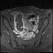
Date of Original Release. Ultrasound of Pelvic Inflammatory Disease. Historically PID has been a clinical diagnosis supplemented with the findings from ultrasonography US or magnetic resonance MR imaging.
Pelvic inflammatory disease PID is a common medical problem with almost 1 million cases diagnosed annually. With permission Download.
Reviewed for content accuracy.
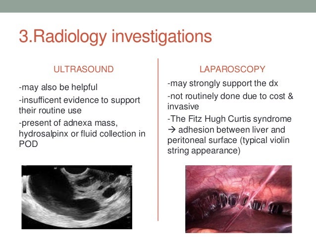
Biopsy which involves taking small tissue samples from the uterus. Pelvic sonography is performed commonly in patients who have a clinical diagnosis of pelvic inflammatory disease. Historically PID has been a clinical diagnosis supplemented with the findings from ultrasonography US or magnetic resonance MR imaging. A right adnexal dead end tubular fluid filled structure with typical mucosal infoldings toward the lumen thick wall and a debris-fluid level ultrasound image characteristic of a dilated fallopian tube in this case. Biopsy which involves taking small tissue samples from the uterus. A connected ultrasound system is another very helpful feature for the PID patient. Ultrasound findings of Pelvic Inflammatory Disease PID. Though PID is clinically diagnosed ultrasound imaging is the first-line radiologic evaluation method of choice for PID and may be used to appreciate the following. 20 21-year-old woman with pelvic inflammatory disease.
Causes of Pelvic Inflammatory Disease. Understanding of the sonographic features of pelvic inflamm. Adnexal mass with heterogenous echogenicity often bilateral Pelvic. Pelvic ultrasound to provide images of the internal organs in the pelvic area. Causes of Pelvic Inflammatory Disease. Livmoder Kvinnligt organ pelvic inflammatory disease stock illustrations. Reviewed for content accuracy.



















Post a Comment for "Pelvic Inflammatory Disease Ultrasound Images"